
Why we care about calcium in the first place
To understand why the calcium flux assay has become such a cornerstone of modern biological research, we first have to appreciate the role of calcium itself. In the world of cellular biology, calcium ions act as a universal language. They are the secondary messengers that tell a heart cell to contract, a neuron to fire, or an immune cell to attack an invading pathogen. Because calcium is involved in so many critical processes, being able to watch it move in real-time is like having a front-row seat to the internal conversations of a living cell.
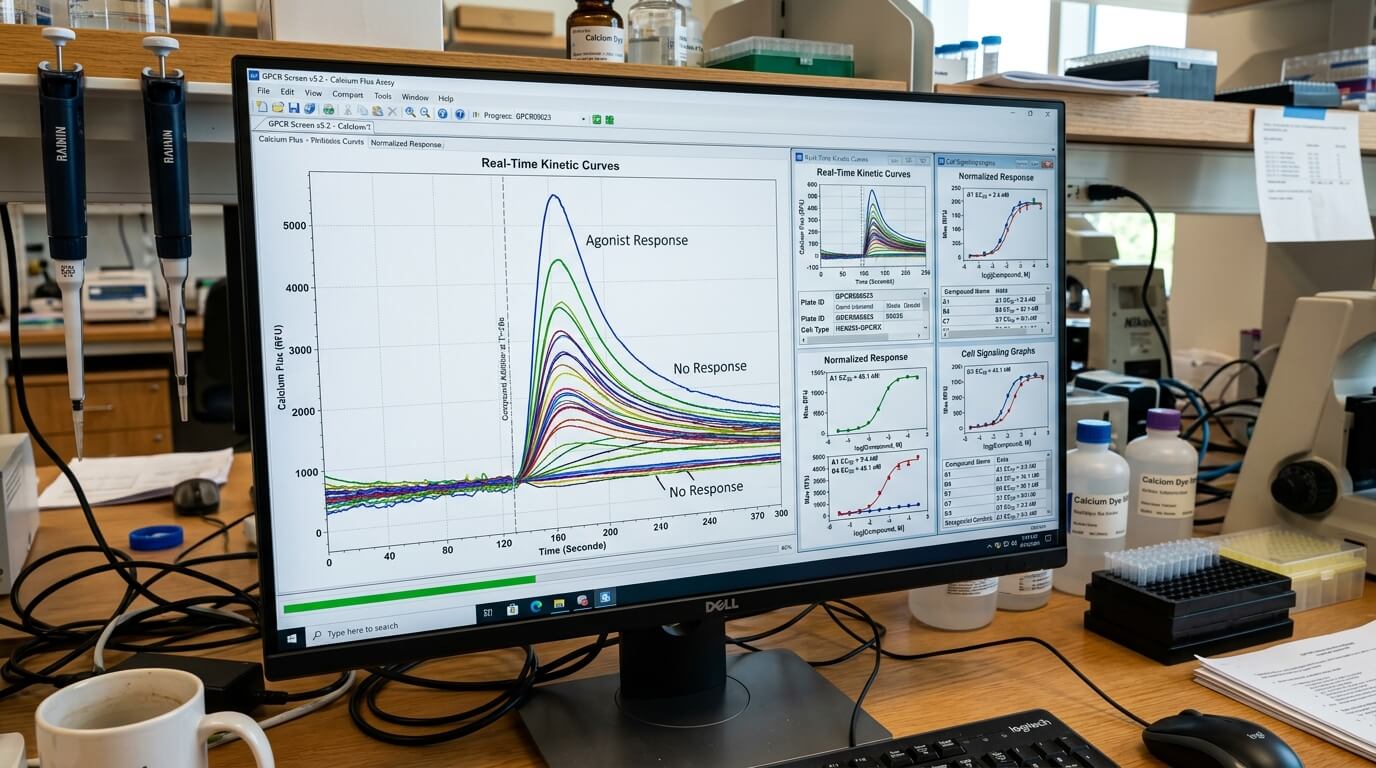
When a cell is stimulated—perhaps by a hormone, a neurotransmitter, or a potential drug candidate—it often responds by rapidly increasing the concentration of intracellular calcium. This ‘flux’ can come from outside the cell through ion channels or be released from internal storage compartments like the endoplasmic reticulum. By measuring this movement, scientists can determine exactly how a cell is reacting to a specific stimulus. This is the fundamental premise of the calcium flux assay, a technique that has been refined over decades to become one of the most reliable tools in the scientist’s toolkit.
The mechanics behind the measurement
At its heart, the assay relies on the use of specialised fluorescent dyes or genetically encoded indicators that change their light-emitting properties when they bind to calcium ions. The process usually begins by ‘loading’ these indicators into the cells. Once inside, the dyes sit quietly until a calcium surge occurs. When the calcium levels rise, the dye binds to the ions and begins to fluoresce more brightly, or in some cases, shifts its emission colour.
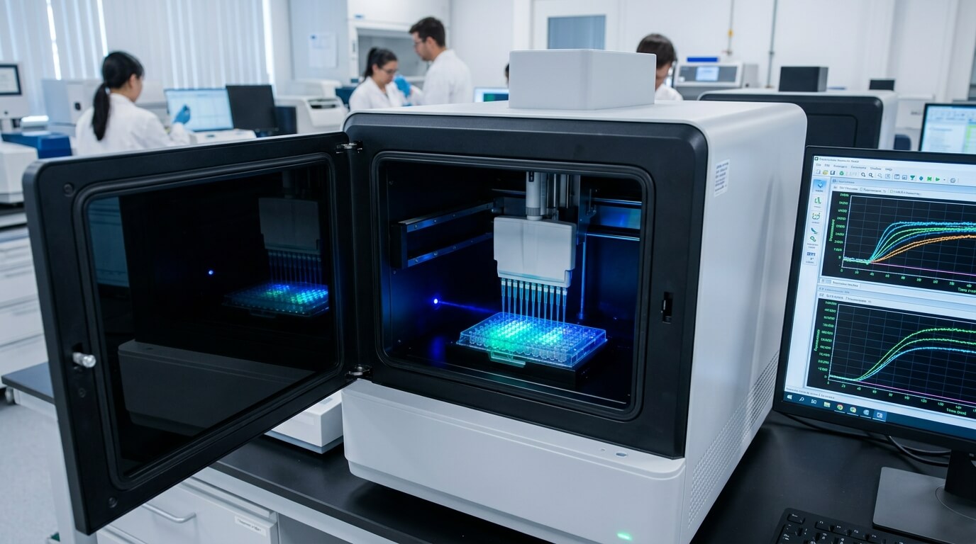
Modern laboratories use sophisticated plate readers and imaging systems to capture this fluorescence. Because these changes happen in milliseconds, the equipment must be capable of high-speed kinetic sampling. This allows researchers to see the entire profile of the calcium response: how quickly it starts, how high it peaks, and how long it takes for the cell to return to its resting state. When pharmaceutical companies are looking for high-quality data to validate new compounds, they often utilise a professional calcium flux assay service to ensure the results are both precise and reproducible.
Key components of a successful assay
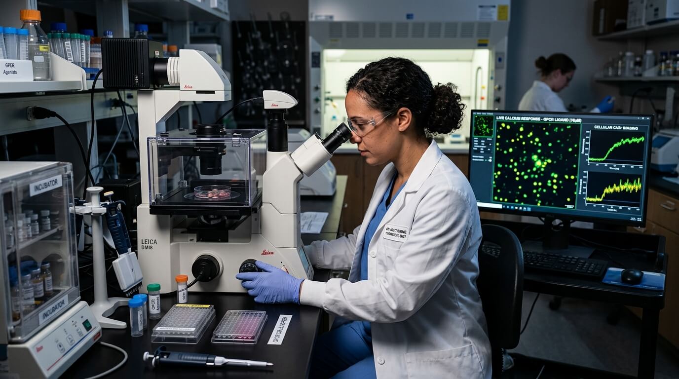
- Fluorescent Indicators: Choosing between ratiometric dyes like Fura-2, which are great for quantifying absolute calcium levels, and non-ratiometric dyes like Fluo-4, which are better for high-throughput screening due to their intense brightness.
- Cell Preparation: Ensuring that cells are at the right confluence and health is vital, as stressed cells often exhibit abnormal calcium signalling behaviour.
- Buffer Optimisation: The extracellular environment must be carefully balanced to prevent background noise while maintaining the physiological relevance of the experiment.
- Detection Systems: High-speed cameras or photomultiplier tubes that can track rapid changes in light intensity across hundreds of samples simultaneously.
Why it is the gold standard for GPCR research
If you look at the top-selling drugs on the market today, a huge percentage of them target G-protein coupled receptors (GPCRs). These receptors sit on the cell surface and act as gatekeepers for external signals. When a ligand binds to a GPCR, it often triggers a signalling cascade that leads directly to a calcium flux. This makes the calcium flux assay an indispensable tool for drug discovery in this area.
The beauty of this assay in GPCR research is its speed and sensitivity. Instead of waiting hours or days to see if a drug has an effect on cell growth or gene expression, researchers can see a response within seconds of adding the compound. This immediate feedback loop allows for the screening of thousands of compounds in a single day, a process known as high-throughput screening (HTS). By identifying which molecules trigger the desired calcium response, scientists can quickly narrow down a library of millions of chemicals to just a handful of promising leads.
Ratiometric vs non-ratiometric approaches
One of the more technical decisions a researcher has to make is which type of dye to use. This isn’t just a matter of preference; it changes the kind of data you get. Non-ratiometric dyes, such as Fluo-8 or Fluo-4, are widely used because they are incredibly bright and easy to work with. They provide a ‘yes or no’ answer: did the calcium level go up? However, they are sensitive to things like dye loading efficiency and cell thickness, which can sometimes make comparisons between different samples tricky.
On the other hand, ratiometric indicators like Fura-2 or Indo-1 are the precision instruments of the calcium world. These dyes are excited by or emit light at two different wavelengths. By taking the ratio of the signals at these two wavelengths, researchers can cancel out variations in dye concentration or cell volume. This provides a much more accurate measurement of the actual concentration of calcium ions, which is essential for detailed physiological studies where the exact magnitude of the signal matters more than just its presence.
Common applications in biotechnology
- Ion Channel Screening: Testing how drugs affect the opening and closing of channels that allow calcium to enter the cell.
- Toxicity Testing: Identifying if a new drug candidate causes unwanted calcium spikes that could lead to cell death or cardiac arrhythmias.
- Neuroscience: Mapping how neurons communicate with one another by observing the calcium waves that travel through neural networks.
- Agonist and Antagonist Profiling: Determining whether a molecule activates a receptor or blocks it from being activated by other signals.
The challenges of getting it right
While the concept sounds simple, executing a perfect calcium flux assay requires a fair amount of finesse. One of the biggest hurdles is background fluorescence. Many of the chemical compounds being tested in drug discovery naturally glow under certain lights, which can mask the signal from the calcium dye. To combat this, scientists use specialised ‘masking’ dyes or wash steps to remove excess indicator from the extracellular space, ensuring that the only light being detected is coming from inside the cells.
Another factor is the ‘leakage’ of dyes. Some cell types have active transport systems that recognise the fluorescent dyes as foreign objects and pump them out of the cell as fast as the researcher can put them in. This requires the use of inhibitors to keep the dye where it belongs. Furthermore, the temperature at which the assay is performed can drastically alter the kinetics of the calcium response. Most physiological processes are optimised for 37 degrees Celsius, but many automated systems operate at room temperature, requiring careful calibration to ensure the data remains relevant to human biology.
Looking toward the future of the assay
As technology advances, the calcium flux assay continues to evolve. We are moving away from simple plate-based measurements and toward high-resolution imaging that allows us to see calcium movement within individual organelles, such as the mitochondria. There is also a growing trend toward using ‘label-free’ technologies or more complex 3D cell models, like organoids, to better mimic the environment of the human body.
Despite these innovations, the core principle remains the same. The ability to monitor calcium flux provides a window into the dynamic life of the cell that few other techniques can match. Whether it is used to discover a life-saving heart medication or to understand the fundamental mysteries of the brain, this assay remains a vital link between chemical stimulation and biological understanding. The integration of automation and artificial intelligence in data analysis is further refined, making the process faster and the insights deeper than ever before, ensuring that this method will remain a staple in laboratories for decades to come.

Chloe is a journalist focused on music genres, artist evolution, and how global platforms have influenced listening habits. She highlights how music connects people across generations.